Adam Lueken

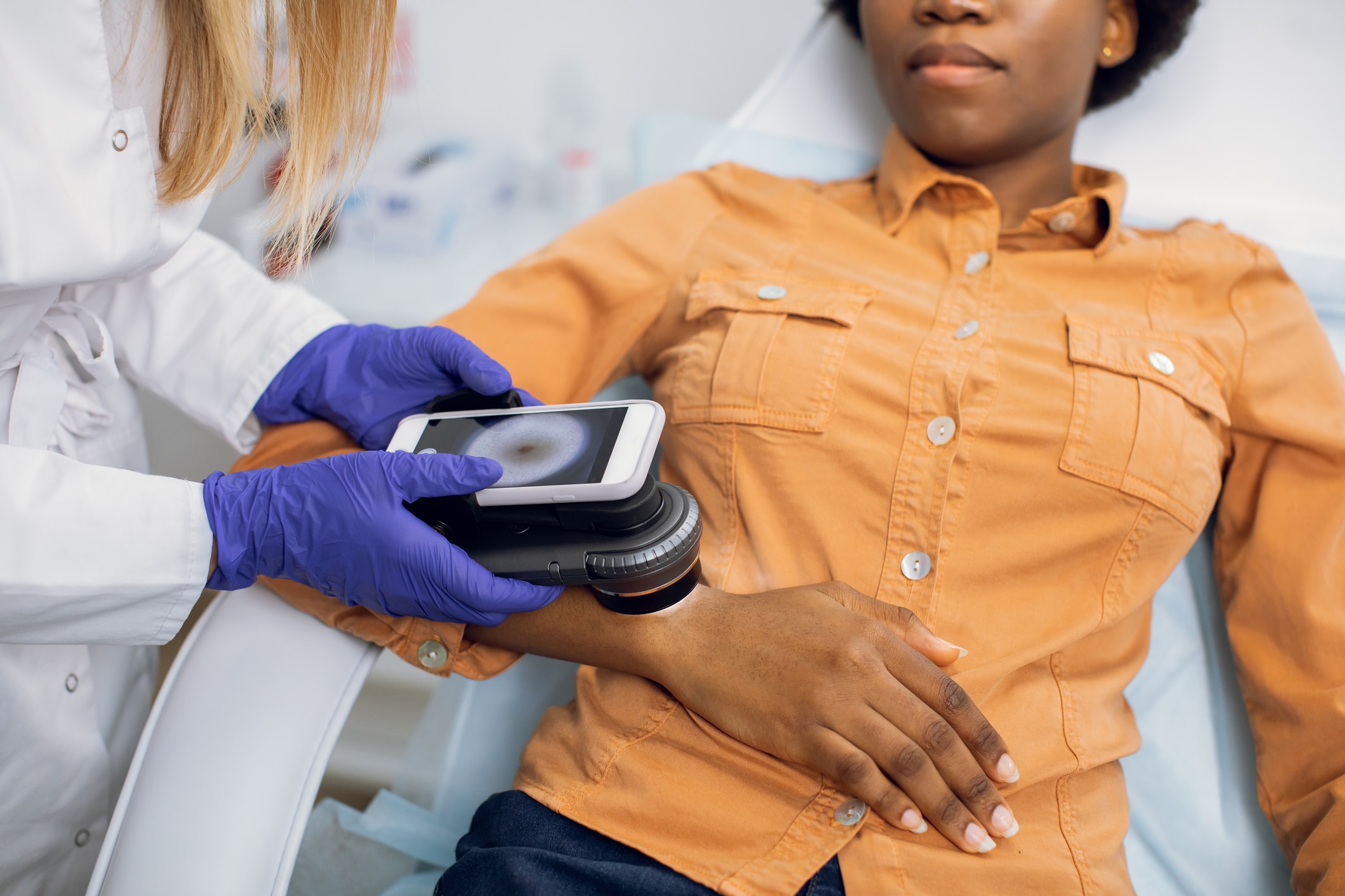
Dermoscopy (or dermatoscopy) is an effective method for evaluating the colors and microstructures of the epidermis, the dermoepidermal junction and the papillary dermis. The process examines cutaneous lesions like BCC (basal cell carcinoma), SCC (squamous cell carcinoma), melanoma, etc. using a dermatoscope. This instrument illuminates and magnifies to show subsurface details. Dermoscopy can also be used to diagnose hair and scalp diseases, nail issues, inflammation and infections.
Many dermatologists use dermatoscopes to identify possible skin problems that a visual examination may not reveal on its own. This can help them discover issues more accurately, which can also reduce the need for more costly and invasive procedures for patients. For skin cancer specifically, it has proven to increase the discovery of malignant melanoma.
If you are not currently using dermoscopy in your practice but are considering making it a part of your skin care, here is some more information on the types of dermatoscopes and how they work.
Polarized and Non-Polarized Dermatoscopes
Polarized devices use polarized light, which creates an accurate visualization of structures located below the superficial dermis, typically with a multispectral or white light. Multispectral light helps dermatologists see things the human eye may not. The technique is based on the specific optical absorption properties of skin pigments. For example, red light can be better for looking deeper into the skin, while blue light may be better suited for more surface-level lesions. A non-polarized dermatoscope requires the use of a contact liquid to cancel out reflections on the skin when making contact. As a result, the device must be cleaned and sanitized after each use, which makes it less convenient. But non-polarized devices are beneficial for identifying certain skin structures that polarized devices cannot.
Analog and Digital Dermatoscopes
Analog dermoscopy involves looking into the magnifying lens for evaluating skin lesions. This process can be done in conjunction with a digital camera for monitoring lesion development over a period of time. Alternatively, digital dermatoscopes (videodermoscopy) have a digital camera built-in, so dermatologists look at the digital screen when evaluating skin instead of a magnifying lens. Digital products also allow dermatologists to take dermoscopic images while examining.
Other Types of Dermatoscopes
There are also hybrid scopes that can be switched from polarized to non-polarized light, and can be used with or without contact liquid. They also feature contact and non-contact modes. Some scopes can also be connected to smartphones to be combined with special add-on lenses. This allows pixel-perfect imaging in some cases. Some dermatoscopes come equipped with software to automate certain imaging processes. And in terms of artificial intelligence, certain dermatoscopes recognize complex skin patterns to help identify signs of malignancy even earlier.
With these different types, there are certainly a variety of options of dermatoscopes, whether digital, analog, polarized, non-polarized or versions that combine the best of all worlds. Depending on your needs, preferences and budget, do some more research on these different options to determine what will work best for you, if jumping into dermoscopy.
Need support implementing new treatments and services? We’re just a quick phone call or click away. Schedule a consultation with one of our practice management experts today!